Experimental diffraction. Evaluating the diffraction pattern
Mode: full-screen / central-screen / help
Table of contents through the logo
To the table of contents
The rotation of a crystal within a monochromatic X-ray beam causes its reciprocal lattice points to cross the surface of the so called Ewald sphere.

Whenever this happens, a diffracted beam is originated in the center of the Ewald sphere and passes through the reciprocal point that lies on the Ewald spherical surface... In these circumstances the so-called Bragg law is fulfilled. The set of all diffracted beams constitute the so-called diffraction pattern, which is subject to detection and evaluation. The reader should be aware that a complete diffraction pattern can contain a highly variable number of diffraction beams, from hundreds (simple inorganic compounds) to hundreds of thousands (proteins or viruses).


Historically speaking, detection of diffracted patterns has been carried out in different ways...

Precession filmWeissenberg photograph
Photographic films showing part of a diffraction pattern
Precession camera (left)  and Weissenberg camera (right)

Four-circle goniometer with a point detector


Four-circle goniometer with an area detector
 Scheme adapted from Carleton College


Independently of the methodology used for the detection of the diffraction pattern, its evaluation and measurement implies three well defined aspects:


Left: Geometric relationship between direct and reciprocal space
Right: Metric relationship between direct and reciprocal space
 

Left: Intensities measured on a photographic film with a photometer
Right: Intensities measured by scanning beam profiles with a point detector


Intensities measured integrating pixels on an area detector


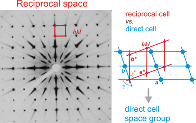
A photographic film showing a reciprocal plane containing the reciprocal points of type hk0. Several possible planes of symmetry, marked with the letter m, are observed.


In short, the end of a full evaluation of the diffraction pattern of a crystal means having obtained a complete description of its reciprocal lattice (geometry + intensities), and hence the knowledge of the direct lattice: unit cell constants (a, b, c, α, β, γ), lattice type (primitive or centered) and crystal symmetry (space group), ie, all ingredients to address the resolution of the internal structure of the crystal.

In general, what has been presented up to this point is enough to understand what the experimental procedures to evaluate the diffraction pattern are (considering that the diffraction pattern contains Bragg peaks only). Therefore, the reader could now go back to the starting point.

However, the advanced reader might take a look below...


How many crystals are needeed and at what temperature is the diffraction experiment done?
 
Obviously, the first diffraction experiments were carried out on stable crystalline materials, such as minerals or inorganic compounds, which are hardly damaged by X-ray radiation, so that one or two crystals were enough to carry out the whole diffraction experiment.

However, later on, crystallographers started dealing with much more labile and complex substances (organic and biological samples) that, due to the rapid deterioration caused by X-ray radiation, required the use of multiple crystals in order to collect their diffraction pattern. As shown in a previous section, this problem was solved through the so-called cryo-crystallography, ie the adaptation of a mechanism for cooling the crystals during their exposure to X-rays, thereby achieving greater sample stability against X-ray damage.


A cryo-protected crystal in an anti-freeze matrix (left), mounted in front of a stream of liquid nitrogen, evaporated at about 100 K (right)

This procedure, still in use for most diffraction experiments in crystallographic laboratories or in synchrotron installations, required the development of a special technique for mounting crystals, using small loops used to "catch" the crystal in a liquid matrix of a cryoprotector (anti-freeze) transparent to X-rays. This procedure is especially relevant for protein crystals. In this type of crystals the cryoprotector matrix is ​​dispersed through their inner channels, replacing the water molecules,  and avoiding their freezing, which would cause the crystal to break. By means of this technique, and also thanks to the high brilliance of the synchrotron radiation sources, the number of crystals needed to carry out a complete diffraction experiment has been greatly reduced, especially in the case of proteins, which usually produce crystals very sensible against radiation and temperature.

However, with the new generations of 
synchrotron and/or XFEL radiation sources, the so-called serial millisecond crystallography is being imposed (Nature Communications (2017) 8, art. 542).The so-called serial millisecond crystallography at a synchrotron beamline equipped with high-viscosity injector and high frame-rate detector allows typical crystallographic experiments to be performed at room-temperature. Using a crystal scanning approach on microcrystals deposited on a grid, one can collect hundreds, thousands, of partial diffraction patterns (the crystal can rotate only few degrees) which can be unified and scaled properly to produce a complete diffraction pattern. Compared with serial data collected at a free-electron laser (XFEL), the synchrotron data are of slightly lower resolution, however fewer diffraction patterns are needed for "de novo" phasing. Overall, the data collected by room-temperature serial crystallography are of comparable quality to cryo-crystallographic data and can be routinely collected at synchrotrons.


Is there anything else besides the Bragg peaks?

 The use of the very powerful radiation sources (synchrotron + XFEL) has led to consider a part of the information contained in the diffraction pattern that was not previously taken into account. We refer to the so-called continuous diffraction, namely an intensity distribution, poorly defined, that may appear around and between the Bragg peaks as well as in areas of higher diffraction angle, where the Bragg peaks have almost disappeared.

The phenomenon of continuous diffraction is negligible in crystals of relatively simple composition, where the dominant aspect is the strict crystalline order and uniformity of molecules. However, it has been shown [Nature (2016) 530, 202-206] that this can be very relevant in the case of biological macromolecules, where the sharpness and intensity of the Bragg peaks can decrease rapidly as a function of diffraction angle (see the diffraction pattern shown in the figure at right).

Although the explanation of these facts goes beyond the scope of these pages, we considered important to add a short explanation of this phenomenon and the importance of its consideration in the world of biological crystallography.

The idea behind the mentioned article is that crystallographic resolution for some macromolecules may be limited not by their heterogeneity, but by a deviation of strict positional ordering of the crystalline lattice. Such molecular displacements from the ideal lattice positions give rise to a continuous diffraction pattern that is equal to the incoherent sum of diffraction from rigid individual molecular complexes aligned along several discrete crystallographic orientations and that, consequently, contains more information than Bragg peaks alone.


Continuous diffraction and coherent diffraction (Bragg)
A still snapshot of the diffraction pattern of a biological macromolecule. The Bragg intensity maxima disapear very quickly with the diffaction angle. In addition, a weak speckle structure is shown beyond the extent of Bragg peaks. Image taken from Nature (2016) 530, 202-206.


Although the existence of continuous diffraction was already known, and it has been used to interpret dynamic phenomena in protein crystals, the above mentioned article provides new important insights for determining the structure of biological macromolecules from diffracting crystals. According to the new considerations, the diffraction intensities contain two terms, one representing the continuous and another one for the Bragg diffraction. The difference is that each term is modulated by different combinations of the asymmetric unit transform. The continuous diffraction is the sum of the intensities of each asymmetric unit transform, whereas the Bragg peaks depend on the coherent sum of the asymmetric unit transforms (the unit-cell transform). See the diagram on the right showing the ocurrence of continuous diffraction versus coherent diffraction.

These considerations imply very significant contributions for macromolecular structure determination, but not just to increase the knowledge of the molecular details (degree of resolution of the model), but also as a tool for assigning phases to diffraction intensities.

Continuous diffraction vs. coherent diffraction (Bragg)
Diagram showing the occurrence of continuous diffraction versus coherent diffraction


But let's go back...
 
Table of contents