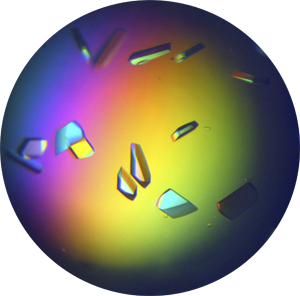
Crystallization is
the process, governed by both thermodynamic and
kinetic factors,
by
which molecules arrange themselves in a natural
manner to form a
repetitive three-dimensional reticulum we
call crystal.
The
crystallization process consists of two major events: nucleation and
crystal growth. Nucleation is the step where the molecules
dispersed in the solvent start to gather into clusters, on the
nanometer scale (elevating solute concentration in a small region),
which becomes stable under the current operating conditions. These
stable clusters constitute the nuclei. However when the clusters are
not stable, they redissolve. Therefore, the clusters need to reach a
critical size in order to become stable nuclei. Such critical size is
dictated by the operating conditions (temperature, supersaturation,
etc.). It is at the stage of nucleation in which the atoms arrange in a
defined and periodic manner which defines the crystal structure
— note that "crystal structure" is a special term that refers
to the relative arrangement of the atoms, not the macroscopic
properties of the crystal (size and shape), although those are a result
of the internal crystal structure.
It is not the purpose of these pages to make a comprehensive
presentation of the different crystallization techniques which can be
found in many textbooks or on different websites, and we will only
refer briefly to some of the most common techniques used to
obtain protein crystals. However, for beginners we recommend to read the brochure prepared
by UNESCO to grow single crystals.
In the case of protein samples, the crystallization experiment begins
with a relatively concentrated
protein solution (between 2 and 50 mg /ml) to which a reagent solution
is added with the intention of reducing its solubility
and generating controlled precipitation. Using a
gradual
increase of concentration and keeping these conditions under control,
tiny crystalline nuclei can be obtained which can grow and
lead to crystals of adequate size for diffraction experiments (between
0.1 and 0.5 mm).


These generic diagrams show the
different areas of a protein-precipitant equilibrium in terms of the
concentrations of both components.
Areas shown in yellow (left) and white (right) represent the conditions
under which the protein is in solution. The areas depicted in blue
represent the conditions under which the protein appears as a
precipitate.
Both areas are separated by another area (shown in pink) with
some supersaturation conditions, suitable for nucleation and crystal
growth.
Thus, to
maximize the possibility of a successful crystallization
experiment, it is necessary to design various experiments, from
different starting positions, ie different concentrations of protein
and precipitant (arrows of different colours in the diagram on the
right).
One of the most common
methodologies for these experiments is based on the hanging
drop technique.
 Scheme of a well reservoir, containing a
precipitant solution, capped with a cover slip, as used in the hanging
drop technique
Scheme of a well reservoir, containing a
precipitant solution, capped with a cover slip, as used in the hanging
drop technique
The procedure consists, approximately, of the following aspects:
A few microliters
(1-2 μl) of protein solution are mixed with
a more or less equal amount of reservoir solution containing the
precipitants (previously
balanced with a pH buffer)
and are deposited on a cover slip which covers the precipitant
reservoir. As
the protein/precipitant mixture in the drop
is less concentrated than the reservoir solution (normally we mix the
protein solution with the reservoir solution at about 1:1), water
evaporates from the drop into the reservoir. As a result the
concentration of both protein and precipitant in the drop slowly
increases, and crystals may form.
On many occasions the sitting
drop technique is also used ...
The market offers several types
of plates suitable for protein
crystallization techniques, as it is shown below. See also the web
pages offered by Hampton Research.

 Left: A 24
well crystallization plate
for protein crystallization using the hanging drop technique
Right: A 24
well crystallization plate
for protein crystallization using the sitting drop technique
Left: A 24
well crystallization plate
for protein crystallization using the hanging drop technique
Right: A 24
well crystallization plate
for protein crystallization using the sitting drop technique
 Left: Under
the right
conditions, crystals can grow in the drop as shown in the picture.
Right: The
available crystallization robots can help to prepare very quickly
hundreds of drops
to find the best initial conditions for crystallization.
Left: Under
the right
conditions, crystals can grow in the drop as shown in the picture.
Right: The
available crystallization robots can help to prepare very quickly
hundreds of drops
to find the best initial conditions for crystallization.
Readers interested in the historical development of
these crystallization methodologies and their influence on
current
practice, should consult the article to be found through
this link.
The animation below
shows the
process where lysozyme crystals are growing from an
aqueous media. The
duration of
the process, that
takes a few seconds on your screen, corresponds approximately to 30
minutes in real life. This case corresponds to an extremely quick
crystal growth process.
With the same
enzyme (lysozyme), the
video produced by Bernhard Rupp shows the relationship between rapid
nucleation and crystal growth rate. The greater the number of nuclei
formed, the lower the growth rate, and therefore the smaller the size
of the crystals obtained.
Compare the size (much
smaller) of the crystals growing in the movie below with
those shown above.
There is a variety of
other
techniques
available to make crystals grow, such as sitting drops, dialysis
buttons, and gel and microbatch techniques.
See also the pictorial
library of crystallization drop phenomena offered by Terese Bergfors at
the University of Uppsala and take a look to this special
issue of Acta Crystallographica.
The advanced reader cannot forget the technical revolution that for
X-ray diffraction has recently been introduced, where
single-crystal
X-ray diffraction ‘snapshots’ are collected from a
fully
hydrated stream of nanocrystals using femtosecond pulses from a
hard-X-ray free-electron laser obtained in a Linac Coherent
Light
Source. This will probably eliminate most "bottleneck" effects
that sometimes crystallization can produce, especially with
proteins (
see this
article published in Nature (2011) 470, 73-77).
But let's go
back...


